

By Patrick Guye, PhD — Co-Founder & CEO, Trilliome
There is a fact about the brain that was taught as settled for most of my working life, and it has just acquired an expiry date.
The immune cells of the brain, called microglia, do not come from your bone marrow. They arrive before you are born, from the yolk sac, in one of the earliest waves of cell migration in embryonic development. They move into the brain, and then they stay. They divide locally to replace their own losses. The blood-borne immune system, the one running everything else in your body, is kept outside.
This was never a casual assumption. Elisa Gomez Perdiguero and colleagues nailed down the yolk-sac origin in a 2014 paper in Nature, using fate-mapping to follow the lineage, and found microglia "only marginally replaced" in one-year-old mice. The following year Julia Bruttger's group ran the opposite experiment. They wiped out microglia in adult mice and watched the compartment refill within a week, entirely from cells already inside the central nervous system, with nothing arriving from bone marrow. Two independent approaches, same answer: the population looks after itself.
So a reasonable person concluded that the immune cells defending your brain at eighty are descendants of cells that arrived before your mother knew she was pregnant.
Human data published this year says that stops being true around fifty.
What the hippocampus shows
Nathan Zemke, Bing Ren and colleagues at UC San Diego profiled post-mortem hippocampal tissue from forty people between twenty and ninety-five years old, none of whom had a neurological diagnosis. They did not stop at which genes were switched on. They mapped gene regulation and the three-dimensional folding of the genome, cell by cell. That combination is what made the finding visible. As Zemke put it, they uncovered a shift in the identity and lineage of immune cells that gene expression alone would not have revealed.
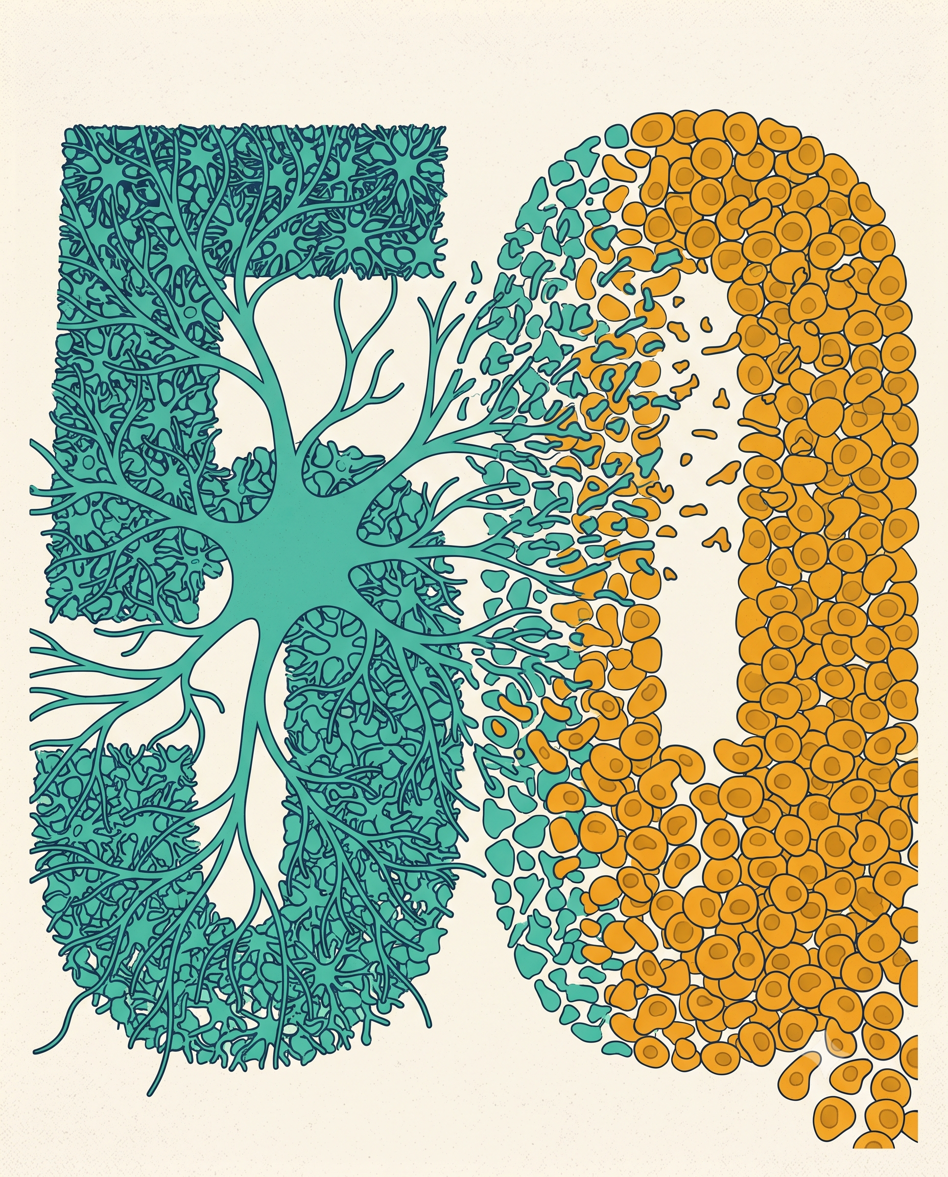
Between roughly fifty and seventy-five, the embryonically derived microglia decline substantially. What takes their place carries the molecular signature of immune cells from the blood.
Two other things happen in the same window. The cells responsible for maintaining the blood-brain barrier deteriorate. And across every cell type they looked at, the three-dimensional architecture of the genome erodes.
Read those together and the picture is uncomfortable in a specific way. The wall thins. The staff changes. Both in people nobody would describe as sick.
The question that opens up
If the aging brain increasingly draws its immune cells from the periphery, then a question that used to be academic becomes practical. Where does the periphery get trained?
A large part of the answer is the gut. And I do not mean that as a figure of speech.
The part I did not expect
In 2020 Zachary Fitzpatrick and colleagues published a study in Nature on the meninges, the membranes wrapping the brain and spinal cord. They found antibody-secreting plasma cells stationed around the dural venous sinuses. Those sinuses are regions of slow blood flow with fenestrations, which in plain terms means they are one of the places where something travelling in your bloodstream has a realistic chance of reaching your brain.
The cells sitting at that vulnerable junction secrete IgA, the antibody class associated with mucosal surfaces. That is already odd for a location inside the skull.
Then they sequenced the B cell receptors and traced the cells back to the intestine.
These are gut-educated cells that have relocated to the brain's most exposed border and taken up guard positions. They are scarce in germ-free mice and return when the animals are recolonised, so their presence depends on the microbiome. When the researchers depleted them or removed IgA, fungal spread into the brain increased after an intravenous challenge. They are doing real defensive work.
And they increase with age.
I have read that paper several times and the detail that stays with me is not the elegance of the trafficking. It is that a routine breach of the intestinal barrier also increased their numbers. The gut is not merely supplying these cells. It is signalling to them.
How the training works
The education itself is chemical, and the mechanism is unusually well established.
When gut bacteria ferment fibre they produce short-chain fatty acids. In 2013 two groups published back to back on what those molecules do to the immune system. Yukihiro Furusawa's team showed that butyrate drives naive T cells toward a regulatory, inflammation-suppressing phenotype, and identified the mechanism: increased histone acetylation at the Foxp3 locus, which loosens the chromatin around the gene that defines those cells. Patrick Smith's group, publishing in Science, showed the same class of molecules controls the size and function of the colonic regulatory T cell pool, acting through the receptor Ffar2.
Both papers now sit above four and a half thousand citations. Mingjing Hu's group extended the finding to human cells in 2022, generating functional human regulatory T cells with butyrate and propionate in the differentiation protocol. That human step matters, because most of this literature is mouse work.
There is a second channel on the same axis, and it runs on nerve. The vagus carries an anti-inflammatory signal from the gut to the brainstem and back out to the immune system, through acetylcholine acting on α7 nicotinic receptors. Population cohorts have linked truncal vagotomy to altered Parkinson's and Alzheimer's risk. I would be careful with that channel, though. A 2022 review by Nicolas Alen went looking for the human evidence behind the cholinergic anti-inflammatory pathway and found substantial gaps between what the mouse work shows and what has actually been demonstrated in people.
And then last week
On 21 July a group led by Grozdan Cvijetic published in Nature Immunology on how a disrupted gut community drives systemic autoimmunity. The initiating event in their model is genetic. Deleting Notch2 in a population of dendritic cells wipes out a subset called cDC2a, and the animals develop an altered gut community, enriched for organisms including Parasutterella and Mucispirillum and depleted of Akkermansia muciniphila and Lactobacillus johnsonii. Those mice expand an inflammatory dendritic cell population, develop autoantibodies, and deposit immune complexes in the kidney.
The genetic lesion is worth naming clearly, because it means this is not a story about diet.
The result that matters is what came next. They transferred the disrupted community into normal mice, with no antibiotic conditioning to clear the way, and reproduced the autoimmune features. The community on its own was sufficient to remodel the peripheral immune system into a pathological state.
What we do not know
Here is where I have to be careful, because the temptation to close this loop is strong.
Nobody has connected the gut to the specific midlife handover Zemke's team described. Not in humans, not in mice. The trafficking route is documented. The chemistry of immune education is documented. The capacity of a gut community to reshape peripheral immunity is documented. The line joining those to a fifty-year-old's changing microglial population is an argument I am making, and the literature has not made it yet.
Two more cautions belong here.
The Zemke work is post-mortem and descriptive. It compares age bands, which establishes that the composition changes, not that the change causes anything. The team says directly that future work is needed to determine whether this immune transition contributes to Alzheimer's disease.
And the incoming cells are not automatically the villains. In a mouse model of Alzheimer's, Ping Yan's group found that monocyte-derived cells made up six percent of plaque-associated macrophages and were reducing plaque load. Removing the spleen cut their numbers and made the pathology worse. Fitzpatrick's gut-derived IgA cells are protective too. A brain that recruits from outside is doing something different at eighty than it did at twenty. Different is not the same as worse.
The question I would want answered
If the composition of your brain's immune system turns over between fifty and seventy-five, and the cells arriving have been educated in a compartment you can actually reach, then the interesting question is one of timing.
An intervention aimed at that window would have to start before the window opens. By the time anyone has a symptom, the handover finished a decade or two earlier. That is an uncomfortable design constraint for a field that mostly runs six-week trials in people who already have a complaint.
It also raises a measurement problem I do not have a good answer to. What would you even track? Nobody is going to biopsy a healthy fifty-year-old's hippocampus to check how the staffing is going.
We spend our time at Trilliome on the upstream end of this, on what specific molecules do to specific bacteria and what those bacteria then do to their host. This particular question sits beyond what anyone can currently claim, mine included. But it is the one I keep coming back to.
If the brain spends its second half hiring immune cells trained somewhere else, the training facility seems worth understanding.
References
- Gomez Perdiguero E, et al. Tissue-resident macrophages originate from yolk-sac-derived erythro-myeloid progenitors. Nature, 2014.
- Bruttger J, et al. Genetic cell ablation reveals clusters of local self-renewing microglia in the mammalian central nervous system. Immunity, 2015.
- Zemke N, Ren B, et al. Epigenetic and 3D genome reprogramming during the aging of the human hippocampus. Science, 2026.
- Fitzpatrick Z, et al. Gut-educated IgA plasma cells defend the meningeal venous sinuses. Nature, 2020.
- Furusawa Y, et al. Commensal microbe-derived butyrate induces the differentiation of colonic regulatory T cells. Nature, 2013.
- Smith PM, et al. The microbial metabolites, short chain fatty acids, regulate colonic Treg cell homeostasis. Science, 2013.
- Hu M, et al. Short-chain fatty acids augment differentiation and function of human induced regulatory T cells. IJMS, 2022.
- Ma L, et al. The vagus nerve: an old but new player in brain-body communication. Brain, Behavior, and Immunity, 2024.
- Alen N. The cholinergic anti-inflammatory pathway in humans: state-of-the-art review and future directions. Neuroscience and Biobehavioral Reviews, 2022.
- Cvijetic G, et al. Dysbiosis-induced expansion of AXL-positive inflammatory type 3 dendritic cells triggers preclinical autoimmunity. Nature Immunology, 2026.
- Yan P, et al. Peripheral monocyte-derived cells counter amyloid plaque pathogenesis in a mouse model of Alzheimer's disease. Journal of Clinical Investigation, 2022.
- Erny D, et al. Host microbiota constantly control maturation and function of microglia in the CNS. Nature Neuroscience, 2015.